“That’s not glass”
Adam Prokai, Indiana University Medical Student // Edited by Michael Barrie, OSU EM Attending.
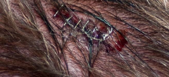
This patient is a middle aged man with a history of alcoholism and a traumatic brain injury resulting in intellectual impairment that required a cranioplasty. The patient presented from an outside clinic for a foreign body that is thought to be glass that had been found in a 3 week old left frontal scalp laceration by nurses one day earlier. The morning of arrival, the patient went to a clinic were a physician saw him, tried to remove the foreign body, stopped, and sent him to the Ohio State Emergency Department. At presentation the patient does not seem to care about the foreign body and describes a history of frequent falls, lacerations, and injuries due to his drinking habits. On physical exam the patient is slow to answer questions and can not give an accurate history sighting multiple blackout episodes and difficulty remembering. A foreign object is found protruding out of the skin where there is a laceration, measuring at least 3cm x 2cm with pus and blood around the object, the object is whitish and opaque. The patient does not remember how long the object has been in the cut or when he first noticed it, but assumes it is from a fall because he “does that a lot and I get hurt.” No other injuries on physical exam, physical exam was normal for all other systems and vital signs were normal.
There are many different patients that are seen in the emergency department every day, one of the most challenging types of patients are poor historians. Without a clear description of symptoms and events, you can not narrow your differential without testing and labs. The goal of medicine is to treat the patient, prevent unnecessary harm and try to not do more testing than indicated. With poor histories it is difficult to do all of these goals at times. So how do you treat someone coming in with an injury and worrisome past medical history who can not give an accurate history?
What Do You Do Next?
The patient is not able to accurately describe the events around this injury and reports differing time frames to multiple providers about when he fell and got the laceration on his head. Basic labs (BMP, CBC), a head CT are ordered to try and visualize the object/injury.
Labs come back normal, and the head CT shows a radiopaque object protruding out of the skin where injury was visualized, and shows an area of fracture over patients cranioplasty site where there is an area missing from the bone graft that matches the size of the foreign object seen on physical exam. It’s about now that as the student I’m glad I didn’t attempt to explore this wound! A neurosurgical consult is ordered and patient is admitted to the hospital for neurosurgical repair the cranioplasty graft.
What to take from this case?
This patient was at an increased risk of injury due to chronic alcohol abuse which has shown to result in greater numbers of traumatic injury when compared to the normal population. The patient is also intellectually impaired due to a previous traumatic brain injury. Patients with intellectual disability have been shown to be at increased risk of fractures and secondary diseases. Both of these factors put our patient at a greater risk for injury and illness when compared to the normal population.
On top of this, the patient has problems fully communicating what his symptoms are and has difficulty giving an adequate history. In order to care for patients with intellectual disabilities, it has been found that having a developed plan in place is the best strategy. Patients with intellectual disabilities may not understand all of their medical problems, and may not be able to communicate what is occurring with words. Using pictures and body movements has been shown to be helpful in communicating in a way that can be better understood. Gaining a history from another source close to the patient can also be helpful to get a clearer story and have the source describe the changes that they have seen in the patient.
In order to adequately treat someone who can not give an accurate history, a plan needs to be developed. This should include looking at other sources for history about the patient, develop techniques besides verbal communication to talk to the patient if they are unable to understand, and do a full work up for all possibilities on a differential in order to make sure that nothing is missed that could cause the patient harm.
The plan for this patient was to perform a history and physical exam, read through previous notes to complete history, and perform labs and imaging. This patient was able to communicate verbally and understood what was going on and extra time was taken to teach the patient about results and what was occurring.
Special thanks to Adam for writing up this interesting case while on an EM visiting student rotation at Ohio State. If you’d like to learn more about being a visiting student at OSU, you can apply via VSAS, more info on the OSU website.
References
- Jamshid Ghajar. (2000). Traumatic brain injury. The Lancet, Volume 356, Issue 9233, Pages 923-929. Retrieved from https://www.sciencedirect.com/science/article/pii/S0140673600026891.
- Lowenstein, S. R., Weissberg, M. P., & Terry, D. (1990). Alcohol Intoxication, Injuries, and Dangerous Behaviors–And the Revolving Emergency Department Door. The Journal of Trauma: Injury, Infection, and Critical Care,30(10), 1252-1258. Retrieved from https://www-ncbi-nlm-nih-gov.proxy.medlib.uits.iu.edu/pubmed/2213933
- M J van Schrojenstein Lantman-de Valk, H., & Noonan Walsh, P. (2009). Managing health problems in people with intellectual disabilities. Bmj,338(Mar20 1). Retrieved from https://www-jstor-org.proxy.ulib.uits.iu.edu/stable/pdf/20511585.pdf?refreqid=excelsior:c7c80b28d165af5bfa1397f9098c4da2.
- Sarmast, A., Andrabi, S., Kirmani, A., & Bhat, A. (2017). Cranioplasty: Indications, procedures, and outcome – An institutional experience. Surgical Neurology International,8(1), 91. Retrieved from https://www.ncbi.nlm.nih.gov/pmc/articles/PMC5461575/.
- Zanaty, M., Chalouhi, N., Starke, R. M., Clark, S. W., Bovenzi, C. D., Saigh, M., Tjoumakaris, S. I. (2015). Complications following cranioplasty: Incidence and predictors in 348 cases. Journal of Neurosurgery,123(1), 182-188. Retrieved from http://thejns.org.proxy.medlib.uits.iu.edu/doi/10.3171/2014.9.JNS14405