Med Student Corner: A Changing Epidemiology – A Patient with Stridor and Sore Throat

By Kelsey Sicker, MD Candidate, Class of 2019 // Edited by Michael Barrie, MD OSU EM Attending

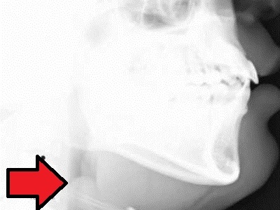
Medicine is a constantly evolving field, with new therapies, preventive strategies, and understandings of disease pathology rapidly growing. As part of this evolution, vaccinations have become key to modern preventive medicine for children and have had a great impact on childhood morbidity and mortality.

With the implementation of universal vaccination recommendations, what long term effects have we seen on the epidemiology of epiglottitis?

Continue reading

Med Student Corner: Heparin Induced Thrombocytopenia

Author: Sandy Brundage (OSU Medical Student Class of 2019) // Editor: Michael Barrie (OSU EM Assistant Professor)

A 56-year old woman presented to the ED for acute onset of shortness of breath with unilateral left leg swelling. PMH significant for a total knee replacement five days ago. Current medications include aspirin, lisinopril, metformin, labetalol and enoxaparin. CBC comes back with a platelet count of 30,000. Upon reviewing her chart, you notice that her platelet count prior to the surgery was 250,000. A lower extremity doppler confirms an acute deep venous thrombosis.

What is contributing to the development of thrombosis despite her medications and thrombocytopenia?

Continue reading

Med student corner: That’s not glass!

“That’s not glass”

Adam Prokai, Indiana University Medical Student // Edited by Michael Barrie, OSU EM Attending.

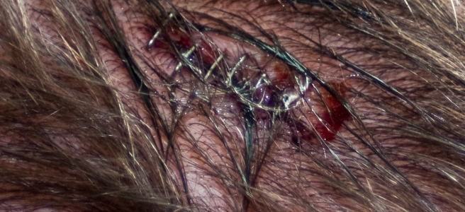
This patient is a middle aged man with a history of alcoholism and a traumatic brain injury resulting in intellectual impairment that required a cranioplasty.  The patient presented from an outside clinic for a foreign body that is thought to be glass that had been found in a 3 week old left frontal scalp laceration by nurses one day earlier.  The morning of arrival, the patient went to a clinic were a physician saw him, tried to remove the foreign body, stopped, and sent him to the Ohio State Emergency Department.  At presentation the patient does not seem to care about the foreign body and describes a history of frequent falls, lacerations, and injuries due to his drinking habits.  On physical exam the patient is slow to answer questions and can not give an accurate history sighting multiple blackout episodes and difficulty remembering.  A foreign object is found protruding out of the skin where there is a laceration, measuring at least 3cm x 2cm with pus and blood around the object, the object is whitish and opaque.  The patient does not remember how long the object has been in the cut or when he first noticed it, but assumes it is from a fall because he “does that a lot and I get hurt.”  No other injuries on physical exam, physical exam was normal for all other systems and vital signs were normal.

There are many different patients that are seen in the emergency department every day, one of the most challenging types of patients are poor historians.  Without a clear description of symptoms and events, you can not narrow your differential without testing and labs.  The goal of medicine is to treat the patient, prevent unnecessary harm and try to not do more testing than indicated.  With poor histories it is difficult to do all of these goals at times.  So how do you treat someone coming in with an injury and worrisome past medical history who can not give an accurate history?

What Do You Do Next?

The patient is not able to accurately describe the events around this injury and reports differing time frames to multiple providers about when he fell and got the laceration on his head.  Basic labs (BMP, CBC), a head CT are ordered to try and visualize the object/injury.

Labs come back normal, and the head CT shows a radiopaque object protruding out of the skin where injury was visualized, and shows an area of fracture over patients cranioplasty site where there is an area missing from the bone graft that matches the size of the foreign object seen on physical exam. It’s about now that as the student I’m glad I didn’t attempt to explore this wound! A neurosurgical consult is ordered and patient is admitted to the hospital for neurosurgical repair the cranioplasty graft.

What to take from this case?

This patient was at an increased risk of injury due to chronic alcohol abuse which has shown to result in greater numbers of traumatic injury when compared to the normal population.  The patient is also intellectually impaired due to a previous traumatic brain injury.  Patients with intellectual disability have been shown to be at increased risk of fractures and secondary diseases.  Both of these factors put our patient at a greater risk for injury and illness when compared to the normal population.

On top of this, the patient has problems fully communicating what his symptoms are and has difficulty giving an adequate history.  In order to care for patients with intellectual disabilities, it has been found that having a developed plan in place is the best strategy.  Patients with intellectual disabilities may not understand all of their medical problems, and may not be able to communicate what is occurring with words.  Using pictures and body movements has been shown to be helpful in communicating in a way that can be better understood.  Gaining a history from another source close to the patient can also be helpful to get a clearer story and have the source describe the changes that they have seen in the patient.

In order to adequately treat someone who can not give an accurate history, a plan needs to be developed.  This should include looking at other sources for history about the patient, develop techniques besides verbal communication to talk to the patient if they are unable to understand, and do a full work up for all possibilities on a differential in order to make sure that nothing is missed that could cause the patient harm.

The plan for this patient was to perform a history and physical exam, read through previous notes to complete history, and perform labs and imaging.  This patient was able to communicate verbally and understood what was going on and extra time was taken to teach the patient about results and what was occurring.

Special thanks to Adam for writing up this interesting case while on an EM visiting student rotation at Ohio State. If you’d like to learn more about being a visiting student at OSU, you can apply via VSAS, more info on the OSU website.

References

Med Student Corner: an atypical presentation flank pain

By Richard Cunningham, OSU M4 // Edited by Michael Barrie, OSU EM Attending

Case:A 54 year old woman with a history of diabetes mellitus type 2, obesity, kidney stones and multiple episodes of pyelonephritis presents with flank pain. It started 3 days ago, has been constant, and intermittently increases in severity. She rates the pain as 4/10. The pain is described as a dull ache, at times sharp, and radiates to the right upper quadrant. The patient endorses some nausea but denies fever, emesis, diarrhea, constipation, chest pain, SOB, dysuria and hematuria. Upon further questioning she says this feels very similar to the times she had kidney infections. She has no history of abdominal surgeries.

Her vital signs are within normal limits and she is afebrile. On exam, she is comfortable appearing and has mild CVA and RUQ tenderness. Her abdomen is soft without guarding or rebound. The rest of her exam is benign.

After reviewing the above case, what is the one lab test (not including imaging) you would order? If you think like me, you would go straight for the UA. Flank pain, history of kidney stones and pyelo, it’s got to be a slam dunk, right? Wrong. Her UA was significant for only trace protein. Well shucks, looks like she’s heading to the CT scanner.

What is your backup diagnosis now? The pain did radiate to her RUQ and she is obese; maybe she has gallbladder pathology? Wrong again…

This woman had appendicitis confirmed on the CTAP.

Atypical Appendicitis:

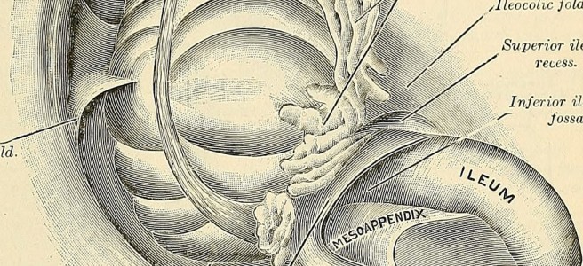
Appendicitis is a common surgical condition traditionally managed with early appendectomy and is associated with low morbidity and mortality. The classic presentation is vague peri-umbilical pain that migrates to the right lower quadrant; this however only accounts for about 50% of patients. The others vary in their presentation due to the age of the patient, the position of the appendix and coexisting conditions such as pregnancy. Retrocecal appendices can be shielded from the anterior abdominal wall by overlying bowel, leading to less severe pain in a non-classic location. A gravid uterus can push the appendix higher into the abdomen, leading to RUQ pain. These atypical presentations can lead to delays in diagnosis and treatment, so it is important to remain vigilant and keep a broad differential.

Some other learning points to consider include being wary of the chief complaint of flank pain. When I see this in the chart I automatically think of the kidney, mainly stones and infection. But it is critical to keep in mind what other pathology can lead to pain in this area and keep a broad differential: low back strain, rib fracture, AAA rupture, zoster of the thoracic dermatomes, pulmonary embolism, cholecystitis, and of course, appendicitis (among others).

Also keep in mind that when the patient tells you when gathering the history that this feels like a condition they have had before, do not let that pigeon hole you into that diagnosis. Stay broad, be creative in constructing your differential. It very well may be a recurrence of that same condition, but sometimes it won’t be and you will get burned. Remember one of the 10 Commandments of Emergency Medicine: Trust No One.

Take home points:

  • 50% of patients with appendicitis presents atypically (it’s normal to not be normal!)
  • Infants and elderly patients are more likely to have atypical presentations
  • Patients with a retrocecal appendix will have more flank pain
  • Pregnant patients will have pain in atypical locations depending on trimester.
  • In flank pain, always keep a broad differential!

References:

Liu CD, McFadden DW. Acute abdomen and appendix. In: Greenfield LJ, et al., eds. Surgery: scientific principles and practice. 2d ed. Philadelphia: Lippincott-Raven, 1997:1246–61.

Ong EMW, Venkatesh SK. Ascending retrocecal appendicitis presenting with right upper abdominal pain: Utility of computed tomography. World Journal of Gastroenterology : WJG. 2009;15(28):3576-3579. doi:10.3748/wjg.15.3576.

https://www.ncbi.nlm.nih.gov/pmc/articles/PMC2715990/
Med Student Corner: Saving Sepsis, Early Recognition and Response

Author: Nisha Crouser, MS4 // Editor: Michael Barrie, OSU EM Attending

One of the first questions Emergency Medicine physicians have to ask themselves is “Do I think this patient is sick or not sick?” A simple question, that takes years of practice and experience to answer. Once a patient is deemed “sick” the questions to follow become more difficult, “How sick?” “What should we do next?” The following case presentation helps elucidate some of the difficulties faced in the ED when sick patients arrive and how to act on the dangerous diagnosis of septic shock.
A 58-year-old patient with several past medical problems including hypertension, hyperlipidemia, and heart disease; presents to the ED with a fever and back pain. Upon arrival the patient is a diaphoretic and ill-appearing obese female who is able to follow commands and answer a few questions. It is difficult to obtain a history from the patient and there is no family present to contribute any information. She is tachycardic and hypotensive when placed on the monitor. Given these vitals in combination with her fever, a Septic Shock alert is initiated. The ED is very busy and there is another severely ill patient that the physician is also covering. Several tests are ordered and fluids are started, there is no obvious source of infection. Over the course of the next half an hour, the internal medicine doctors lay eyes on the patient who has now become more unresponsive. As another half an hour goes by, the patient’s skin become extremely mottled and blue. Her oxygen saturation and blood pressure drop precipitously and she goes into asystole.

Continue reading

Med Student Corner: My Arm’s on Fire! Or the Clever Masquerade of Cellulitis

Author: Lilamarie Moko, MS4 // Editor: Michael Barrie, OSU EM Attending

It’s another late Thursday night in the ED and the waiting room is backed up with patients needing care. As you finish up with your seventh patient presenting with undifferentiated abdominal pain, your eyes rove the board for something new.  You assign yourself to a gentleman in his 60s, a “John Doe” with left arm swelling. Hopefully his abdomen has been behaving itself…

Upon entering the patient room, you see a pleasant, comfortable appearing gentleman in his mid-60s. He’s been having left arm pain and swelling that started 6 days ago. His arm is “on fire”. He first noticed some redness in his left elbow, associated swelling, tenderness, and restriction flexion and extension of his elbow. After about a day, he noticed the redness spreading down to his left wrist, with similar swelling, tenderness, and restricted flexion and extension of both wrist and fingers. He’s had a temperature to 100F, diaphoresis and chills. Also he reports some diarrhea, and urinary frequency in the past 2 days. With some further probing, he reveals that he just returned from Florida several days ago, had no noted scrapes or wounds, and spent most of his time fishing. He works as a farmer, but hasn’t been working for the past couple weeks.

Continue reading

Med Student Corner: Ankle Pain – Reviewing the Ottawa Ankle Rules

Chief Complaint: Ankle Injury–Does this patient have a fracture?

Author: David Sacolick, MS4 // Editor: Michael Barrie, OSU EM Attending

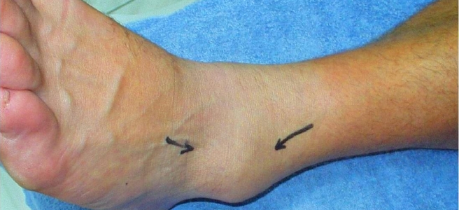
Musculoskeletal injuries are common chief complaints in both emergency medicine and primary care settings. In particular, over 5 million ankle injuries occur in the United States each year. This patient population includes both young active patients as well as elderly patients. And while ligamentous sprains are more common, fractures are also common and can have long term consequences if not appropriately treated.

When a patient presents with a chief complaint of an ankle injury, how do you answer the question: Does this patient have a fracture?  Continue reading

EKGs and Chemistries: AMP Rapid Fire Case Conference Review, November 29, 2017

Welcome back to another week of rapid fire case conference review here at THE OSU EM Residency Program.

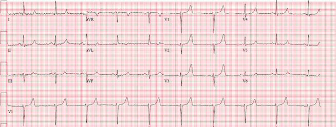
Leading off is Dr. Nicholson with his patient presenting from dialysis clinic with a chief complaint of “Problem with fistula site.” A quick duplex study with basic labs reveals an occluded outflow vein and a potassium of 7.3. Understanding the arrhythmogenic potential for hyperkalemia, he obtains the following EKG:

Screen Shot 2017-12-17 at 8.24.49 PM.png

Continue reading

Fussy Neonate – a Jeopardy game!

Maya S. Iyer, MD, FAAP, Clinical Assistant Professor of Pediatrics, Nationwide Children’s Hospital, Section of Emergency Medicine

Fussy Neonate Jeopardy

This jeopardy game highlights key clinical considerations for infants who present to the emergency department with the chief complaint of fussiness. In particular, the topics highlighted in this game include: fever in the neonate, sudden infant death syndrome (SIDS), brief resolved unexplained events (BRUE) and apparent life threatening events (ALTE), non-accidental trauma (NAT) and a potpourri of other interesting clinical conditions.  The questions require second order thinking. After completing this game, emergency medicine residents should be able to describe the cardinal signs and symptoms, management and possible complications of the above conditions.


Benzos, Bleeding, Burns. Case Conference Review, November 8, 2017


Welcome back to another edition of Case Conference Review here at Academic Medicine Pearls at THE Ohio State University! Old Man Adams starts us off with a 38-year-old male with known history of alcohol abuse presenting via EMS for suspected EtOH withdrawal. On walking into the room, Dr. Adams is greeted with choice expletives and the subsequently refuses any vitals or to participate in the examination. The patient then promptly starts to seize, sending Dr. Adams down his alcoholic withdrawal seizure pathway.

Continue reading